Fetal development is the term used to refer to human development before birth. Human development begins at conception or fertilization and continues throughout the individual’s lifetime. Conception or fertilization refers to “the process by which the male’s sperm unites with the female’s ovum. By this event, a new life is created and the sex and other biological traits are determined. These traits are determined by the combined genes and chromosomes that exist in the sperm and ovum.”1
The new life that begins at conception/fertilization is referred to by different terms depending on the stage of its development. From conception until its implantation into the uterus, the new life is referred to as the zygote. Once implantation occurs between 7-10 days after conception, the growing life is known as the embryo. The embryonic stage of development continues until week eight, at which point all the organ systems are present, the heart is fully developed and the embryo looks like a tiny human being. From week eight until birth, the life in the womb is referred to as the fetus.2 During the fetal phase, the organ systems become more mature, and the fetus grows from approximately 3cm and 2.5g at 60 days to 50 cm and 3300g at term.3
During the zygotic and embryonic stages, development occurs by cell division and specialization into various organ systems. The fetal phase is characterized by maturation of the existing organs and immense fetal growth. 4
Life Begins (Weeks 1 and 2)
The moment the ovum and sperm join at conception/fertilization, the life of a new, distinct, human individual begins. Fertilization of the ovum usually occurs as it travels from the female ovary through the uterine tube, and into the body of the uterus. When the sperm and ovum unite, the resulting zygote develops from one cell into about 32 cells by day four after fertilization. By this time, the cells start to specialize, either into embryonic cells, or cells that will form the placenta and the membranes that will surround the embryo. Within 72 hours of fertilization, the zygote reaches the uterus where it will implant 7-10 days after fertilization.5 By the end of the second week, implantation is complete.6,7
As implantation occurs, the placenta starts to form. The placenta is an essential, though temporary organ, which serves as a vital pathway between the mother and fetus. It transfers oxygen from mother to fetus, and carbon dioxide from fetus to mother. It transports nutrients from mother to fetus, and wastes from fetus to mother. It transfers heat from mother to fetus, and helps to regulate the effect that maternal drugs and medications have on the fetus. It produces hormones essential for fetal growth and for the continuation of the pregnancy.8, 9
Week 3
During the third week, the embryo’s heart and circulatory system,10 kidneys,11 brain, spinal cord, gastrointestinal tract and diaphragm12 start to develop. Leg and arm buds begin to form13 and the embryo is approximately 2 mm long from crown to rump.14
Week 4
By week four, the embryo’s leg and arm buds become more noticeable. The lungs begin formation;15 the liver and pancreas appear as buds16 and a “single-tubed heart begins to beat at 21 – 25 days from fertilization.”17 By twenty-eight days’ gestation, the heart is able to circulate its own blood.18
Week 5
By the fifth week, the leg and arms buds are more fully developed.19 The eyes and ears begin to form and nerves become visible in the brain.20 It is during the fifth week that the brain, heart and circulatory system will “show the most advanced development.”21
Week 6
The sixth week involves development of the external ears,22 primitive skeleton and central nervous system. Brain waves can be detected, and the liver begins to produce red blood cells.23 At this stage “the upper and lower jaws are recognizable … the upper lip has formed, and the palate is developing.”24
|
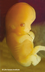
Seven week old embryo |
Week 7 During week seven, the arms and legs begin to move, elbows and toes are visible and the mouth and lips become visible. Teeth buds, hair follicles, the diaphragm25 and nipples begin to form.26 The eyelids and tongue are also starting to develop.27 Towards the end of the seventh week, cartilage will begin to develop, and this cartilage will soon start converting into bone.28 |
| Week 8 By week eight, the embryo’s heart is complete and facial features continue to develop.29 External genitals begin to form according to the gender determined at conception. During the eighth week, the larger muscles are able to contract and bones start to form. By the end of this week, all the major organ systems are present,30 the embryo “clearly resembles a human being” and is approximately 3 cm long from the crown of the head to the rump.31 This marks the end of the embryonic stage of development. |
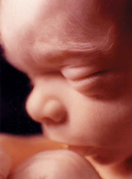
 Eight week old embryo. Eight week old embryo.At this stage of development the embryo looks like a tiny human being. |
|
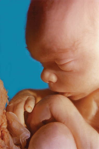
11 week old fetus |
Week 9 – 12 The beginning of week nine signals the start of the fetal stage of development. Between the ninth and twelfth week of development the head will account for almost half the size of the fetus. The digestive system begins to function, urine is produced and excreted, blood starts to form in the bone marrow,32 vocal cords begin to form allowing the fetus to make sounds33 and the face, neck, fingers and toes are almost completely formed. By twelve weeks’ gestation the fetus can make a fist, weighs approximately 45 grams, is 5 cm long from crown to rump and has a heart beat that can be detected electronically.34 At this point in development the gender of the fetus can also be determined visually.35 |

| Week 13 – 16 During week thirteen to sixteen, fine hair develops on the head, bones become harder, the fetus starts to make sucking motions, the skin is transparent, and fingernails are detected. By week fifteen, the sensory system begins to function, allowing the fetus to sense pressure and touch, as well as feel pain.36 The fetus will also gain 4 times its weight during these four weeks.37 The mother may be able to feel fetal movement by the sixteenth week.38 By the end of the sixteenth week the fetus will have a functioning pancreas and liver and will also be able to swallow amniotic fluid.39 |
|
|
|
Week 17 – 20 By the seventeenth and eighteenth week of development, the heartbeat can be heard with a stethoscope. The eyebrows and head hair appear, the muscles are well developed40 and the fetus can produce antibodies. By the end of the twentieth week the fetus will have developed “definite sleeping and activity patterns … that will guide sleep/wake patterns throughout life.”41 By the twentieth week, the fetus weighs 435 – 465 grams and is approximately 19 cm long.42 |
Week 21 – 24
Between weeks twenty-one and twenty-four, the eyelashes and eyebrows are almost fully formed, and the pupils react to light. The fetus responds to loud or sudden sounds with a startle reflex and can grasp with his/her hands.43 The formation of blood in the bone marrow increases as formation of blood decreases in the liver.44 At this stage in development the fetus has reached viability, which means that it has the chance of surviving outside the womb.45 At twenty-four weeks’ gestation the fetus weighs approximately 780 grams and measures 24 cm from crown to rump.
| Week 25 – 28 During weeks twenty-five to twenty-eight, the formation of blood cells completely transfers to the bone marrow, the central nervous system controls some functioning, the eyelids can open and close and the fetus has its own distinct fingerprints. The fetus is now approx. 28 cm long, weighs about 1200-1250 grams and usually moves into a head-down position to prepare for birth. |
|
Week 29 – 32
Between the twenty-ninth and thirty-second week, the central nervous system takes more control of body functions and the rhythmic movement of breathing begins. The lungs are not yet fully mature at this stage. By the end of the thirty-second week, the fetus responds to sounds from the outside world.46 Body fat rapidly increases, and by the end of week thirty-two, the fetus weighs almost 2000 grams47 and measures 30 cm.48
Week 33 – Term
From week thirty-three until birth, body fat continues to increase, fine hair starts to disappear and breast buds form on males and females. The mother supplies the fetus with antibodies against disease and the fetus completely fills the uterus. The final weeks of development are committed to the maturation and growth of the fetus. During the final weeks, the fetus gains approximately 14 grams of fat a day.49 All body systems are functioning, and the fetus is ready to meet the world outside the mother’s womb. Given oxygen, nutrition, and a nurturing environment, the healthy fetus will continue to develop into an infant, toddler, adolescent, adult, and senior, until death.
References
1 Miller BF, et al. Encyclopedia and Dictionary of Medical, Nursing and Allied Health, 5th Edition. W. B. Saunders Company, 1992:547.
2 Pillitteri P. “Maternal & child health nursing: care of the childbearing & childrearing family – 5th edition.” Lippincott Williams & Wilkins: Philadelphia, 2007: 183.
3 Seeley RR, et al. “Anatomy and Physiology, 2nd Edition. Mosby.” Year Book Inc: St Louis Missouri, 1992: 948-66.
4 Gardosi J. “Normal fetal growth,” in Dewhurst’s textbook of obstetrics and gynaecology – 7th edition. Edited by D. Keith Edmonds. Blackwell Publishing: Massachusetts, 2007: 30.
5 Pillitteri. p. 184.
6 Ricci SS. “Essentials of maternity, newborn and women’s health nursing.” Lippincott Williams & Wilkins: Philadelphia, 2007: 212.
7 Vidhya T, et al. “Normal embryonic and fetal development,” in Clinical Obstetrics: the fetus & mother handbook. Edited by E. Albert Reece, et al. Blackwell Publishing Ltd.: Massachusetts, 2007.
8 Lotas, MJ, et al. “Critical Periods in Development” in Developmental care of newborns & infants: a guide for health professionals. Edited by Carole Kenner, et al. Mosby: Philadelphia, 2004: 90.
9 Reeder, S., et al. “Maternity Nursing: Family, Newborn and Women’s Health Care,17th editon.” J.B. Lippincott company, 1992:155-162.
10 Vidhya. et al.
11 New Jersey, 2004:239
12 Seeley. et al. p. 958-959.
13 Ricci. 316.
14 Olds. et al. 239.
15 Vidhya. et al.
16 Seeley. et al. p. 958-959
17 Seeley. et al. p.958-959.
18 Olds. et al. p. 239
19 Olds, et al. p. 239
20 Ricci. p. 216.
21 Olds. et al. p. 242.
22 Jones RE. “Human reproductive biology – 3rd edition.” Elsevier Inc.: Massachusetts, 2006: 10.
23 Ricci. p. 216.
24 Olds, et al. p. 239.
25 “Midwifery by ten teachers.” Edited by Debbie Holmes, et al. Oxford University Press: New York, 2006: 21.
26 Ricci. p. 216.
27 Olds, et al. p. 239.
28 Cronin A, et al. “Human Development and Performance Throughout the Lifespan, 1st edition.” Thomson Delmar Learning: New York, 2005: 105.
29 Ricci. p. 216.
30 Seeley. et al. p. 948.
31 Olds. et al. p. 242.
32 “Maternity Nursing, 7th edition.” Edited by Lowdermilk DL, et al. Mosby Inc.: Missouri, 2006: 202.
33 Lotas, et al. p. 99.
34 Olds. et al. p. 243.
35 Ricci. p. 216.
36 Lotas. et al. p. 99.
37 Ricci. p. 216.
38 Cronin. et al. p. 108.
39 Pillitteri. p. 196.
40 Ricci. p. 216.
41 Pillitteri. p. 196.
42 Olds. et al. p. 243.
43 Pillitteri. p. 196.
44 “Maternity Nursing.” p. 203.
45 Cronin. et al. p. 108.
46 Pillitteri. p. 196.
47 Olds. et al. p. 244.
48 Cronin. et al. p. 96.
49 Cronin. et al. p. 110.